|
Choose: |
|
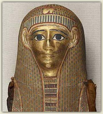
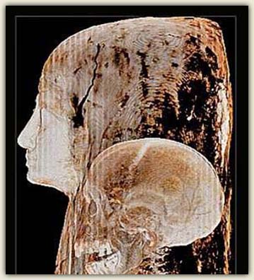
Place your mouse over 1 and 2 to see how CT scans allow us to see through caskets to view the mummy lying within. |
|
Tomography comes from the Greek word tomos which means "slice". This analysis is used to virtually cut an object and see inside it using penetrating radiations, such as X-rays, gamma rays, neutrons, muons and ultrasounds.
CT allows to “see inside” artworks and therefore, to learn about the artwork in a non-invasive manner. CT is able to produce an interior detailed image, very useful for a 3D reconstruction of the object. CT is more and more used for the conservation and restoration in the cultural heritage field because this kind of analysis can help to understand the construction techniques and the history of the object under examination. While CT is successful for the analysis of mummies, statues, vessels and other little objects, CT is not the proper technique to detect paint layers in a painting. It is more useful for the localization of other bigger structures over or under the painting’s surface.
A mummy with serious dental problems
Mummies are encased in their elaborately decorated linen and plaster cases and, for a number of reasons, can’t be taken out for the purpose of scholarly and scientific research.
Are the secrets of mummies forever forbidden? Is there a non-invasive way to learn more about mummies?
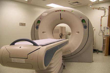
Thanks to tomography, there are ways that modern science can reveal the secrets of mummies without disturbing the corpse or the burial case. By running a CT scan of the mummy in a medical facility as if it were a patient, you can uncover what lies inside the ceremonial burial case.
Looking at the tomographic slices you can assert that this mummy was a woman between 50 and 60 years old and she was quite small. You can also dig up a great amount of other details about her life. You can understand that she suffered from severe dental problems, including at least sixteen abscesses, as well as one tooth that had a dental prosthesis that was probably made from resin.
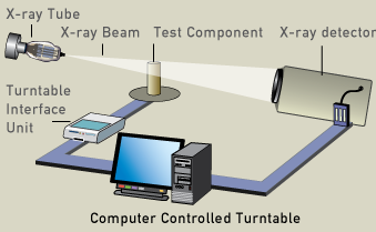
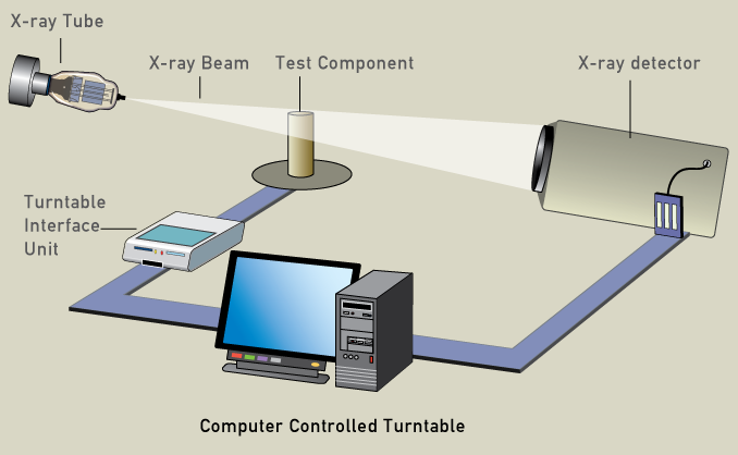
Computer controlled turntable:
Inside the CT scanner there is an x-ray tube and an x-ray detector on the opposite direction which can record the amount of x-ray passing through an object between the x-ray tube and the detector. The sample is moved by means of a turntable around one of its axis and many x-ray pictures of the same area are taken from many angles. This data is then transmitted to a computer, which builds up a 3-D cross-sectional picture of the object and displays it on the screen.
How it works
X-ray computed tomography is based on the absorption of x-rays crossing the object. Tomography is the process of generating a two-dimensional image of a slice or section through a 3-dimensional object (a tomogram). The CT scanner uses digital geometry processing to generate a 3-dimensional (3-D) image of the inside of an object. The 3-D image is made after many 2-dimensional (2-D) x-ray images are taken around a single axis of rotation - in other words, many pictures of the same area are taken from many angles and then placed together to produce a 3-D image. Inside the CT scanner there is an x-ray tube and an x-ray detector which can see hundreds of different levels of density. This data is transmitted to a computer, which builds up a 3-D cross-sectional picture of the part of the body and displays it on the screen.
Technical details
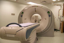
CT systems are available in hospitals, in some universities and in some firms working in the field of non-destructive evaluations. CT systems in hospital are the best devices to analyze mummies because they are already optimized for objects in the shape of human bodies.