Peripheral Vision
We have surprisingly low visual acuity (resolution) in parts of the visual field that are not at the center of gaze — where we are looking. We are not aware of this because we instinctively direct our center of gaze to where we are looking.
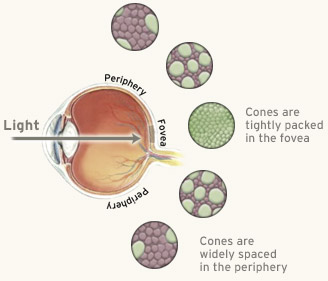
The center of gaze, called the fovea, has a higher density of cones than anywhere else on the retina. In fact, at the fovea, there are no rods at all. (In the diagram at right, the cones are shown in green.) The fovea evolved to have the highest possible visual acuity, and the cones are as small as they can possibly be and still function. Moreover, in the fovea, the retinal ganglion cells have smaller receptive fields, and in the periphery, they have much larger receptive fields.
Curiously, despite the vitality of cones to our vision, we have 125 million rods and only 6 million cones.
The density of cones in our center of gaze is shown in the graph above. The peak is on our fovea. The edges of the graph are our peripheral vision.
The fact that our vision has the highest acuity in the center of gaze does not mean that our vision in the rest of the visual field is inferior — it’s simply used for different things. Foveal vision is used for scrutinizing highly detailed objects, whereas peripheral vision is used for organizing the broad spatial scene and for seeing large objects. Our foveal vision is optimized for fine details, and our peripheral vision is optimized for coarser information.